
Among the diseases of dogs are those that cannot be attributed to particularly dangerous, but still, they deliver a lot of inconvenience and discomfort to the pet, for example, hyperkeratosis. This is an excessive thickening of the stratum corneum of the epidermis. One of the mechanisms of skin protection is the process of keratinization or keratinization. Normally, the cells of the surface layers of the skin gradually accumulate the keratin substance, lose their core and turn into a dry, dense scale. Gradually, the skin is getting denser; it cracks, it hurts. For solutions regarding hyperkeratosis dog paw the following information are important.
Nasodigital hyperkeratosis
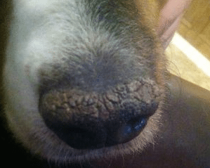
This disease is characterized by excessive keratin formation on the fingertips and nasal mirror. Nasodigital hyperkeratosis can be idiopathic, secondary to an underlying disease, and also observed in congenital disorders of the normal anatomical structure of the nose and fingertips. The idiopathic form of nasodigital hyperkeratosis develops for unknown reasons and is more often noted in the elderly animal. In the case of congenital abnormalities of the anatomical structure, contact of the affected area with the rubbing surfaces occurs.

So, in case of anatomical disturbance of the structure of the nose, the dorsal parts of the nose are not subject to friction during eating, as a result of which the excess keratin is not removed properly. Approximately the same thing happens with the finger pads: in animals with anatomical disturbance, part of the pads does not touch the rubbing surfaces, and excessive keratin deposition is formed.
Thick skin or hyperkeratosis:
Nasodigital hyperkeratosis secondary to the main diseases can be observed with hereditary disorders of keratinization processes, infectious diseases, congenital food dermatoses, dermatosis responding to zinc therapy, necrolytic migrating erythema, skin lymphoma, familial hyperkeratosis of the finger pads.
Clinical signs
In the case of senile idiopathic nasodigital hyperkeratosis, excessive keratin is formed on the nasal mirror and fingertips with a large variety of external manifestations in each individual animal. Usually, an excess of keratin protrudes on the dorsal surface of the nasal mirror and along the edges of the finger pads. The nasal mirror itself becomes dry, rough and hyperkeratotic; the fingertips are affected in a similar way. With significant damage, the formation of cracks, erosions, ulcers and pain is likely.
When nasodigital hyperkeratosis due to violations of the anatomical structure is observed then the formation of an excess of keratin layer in places of reduced contact of the nose and fingertips with rubbing surfaces is required. When hyperkeratosis is secondary to any disease, in addition to thickening of the paws and nose, there are signs of the underlying disease.
The diagnosis
In cases of idiopathic suprasidigital hyperkeratosis and hyperkeratosis in case of congenital anatomical disorders, the diagnosis is made on the basis of characteristic clinical manifestations and usually does not cause difficulties. When hyperkeratosis is secondary to the underlying disease, it is identified by various diagnostic tests. Histopathological changes in the idiopathic form of the disease reveal hyperplasia of the epidermis with severe orthokeratotic or parakeratotic hyperkeratosis in the absence of other changes characteristic of primary diseases.
Treatment
The intensity of treatment depends on the severity of the lesions. Tear ducts should be flushed. Due to the fact that the deposition of excess keratin cannot be stopped, therapy is aimed at its softening and removal. Treatment also depends on the severity of the lesion and the development of secondary complications such as cracks, erosions and ulcers.